Suite 12, 38 Meadowvale Avenue Sth Perth

Complete skin examination for a risk free life | LEARN MORE HERE.

MOLES OR NAEVI

Naevi are benign growths of melanocytes which are the cells that produce melanin, the pigment in the skin.

What causes naevi?

The critical events which lead to the development of naevi remain a mystery. Exposure to ultraviolet radiation (UVR) and genetic factors both play a role. There appears to be a variable relationship between skin, eye and hair colour, the tendency to sunburn and freckle and the development of naevi.

When do naevi appear?

The majority develop throughout the second and third decades of life although some may appear in the first 6 – 12 months of life. Not uncommonly, naevi may also be present at birth (congenital naevi) but these seem to be different from those acquired after birth. They occur equally in both males and females.

What happens to naevi in the course of a lifetime?

Generally an indivudual naevus grows slowly over many years and ultimately attains its full size and then remains stable for decades. Eventually many naevi regress such that very few true moles are present in the 7th and 8th decades.

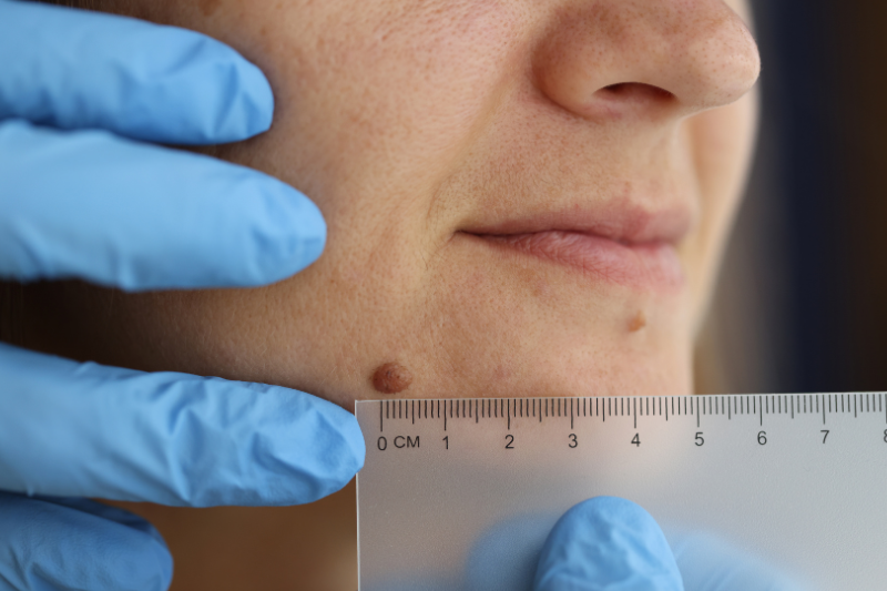
As we age, new naevi are rare and that is why a growing lesion in an adult has a greater risk of being a melanoma.

Sometimes the body’s own immune system “attacks” a mole. An area of depigementation (whitening) occurs around the mole and ultimately the mole may disappear. This clinical picture of a mole with a surrounding pale ring is termed “halo naevus”.

There is a huge range as to what is considered a normal number of moles.  In studies of fair skinned Australians, in the third decade of life, the average number of moles per person is 35.

No. Appearances vary considerably but in general they appear “orderly” with an even surface and colour, are round or oval in shape with regualr edges and relatively sharp borders. They may be dome shaped or flat topped, fully flat or warty or fleshy or appear to be growing on a stalk. They may be flesh coloured, pink or brown. More elevated naevi tend to more lightly pigmented, flatter naevi tend to be darker in colour. Very dark borwn and black are unusual colours of naevi in lightly coloured skins, but are common colours in darkly pigmented skins. Blue – grey, red and white in naevi are not typical and ought to be viewed with suspicion. Naevi may grow hair which may be coarser, longer and darker than in surrounding skin.
Multiple studies demonstrate an increased melanoma risk in individuals with large numbers of naevi. For example in patients with 100 or more naevi, there is a 3 times greater risk compared with those with fewer that 25 naevi. This relative risk was increased to 12 times for patients with 10 or more dysplastic naevi. In patients with numerous naevi (and/or dysplastic naevi) the naevi themselves are best viewed as a marker of increased melanoma risk, not as precursors or pre-cancers of melanoma (see below).
The annual risk of a melanoma developing in an individual mole has been calculated to be approximately 1 in 200,000. Additionally, it seems that only one quarter of melanomas are found associated with a naevus, so that it appears that the majority of melanomas develop directly from “normal” skin.
Absolutely not, because the vast number of moles are harmless. Patients with numerous naevi, especially dysplastic naevi and a personal or family history of melanoma should be screened regularly for life. Moles may be treated for cosmetic reasons, because they are being recurrently traumatised or because there are suspicious features. In the latter scenario, complete excision is recommended so that full microscopic analysis can be performed. Clincially suspicious moles must never be treated with liquid nitrogen, diathermy, dermabrasion or laser. It is acceptable to shave clincally benign lesions for cosmetic reasons as this procedure is associated with much less scarring. However, a pathology check is always advised.
Possibly, but only through a comprehensive sun protection program commencing in early childhood.